Anatomy of the portal system :
The portal vein is formed behind the neck of pancreas, at the level of L2, by the superior mesenteric and splenic veins.
It ascends up along the free edge of lesser omentum, behind the common bile duct.
It enters the liver by dividing into two of it's tributaries.
The left and right gastric veins joins to it.
The inferior mesenteric veins drains into the splenic veins.
Portal vein is valveless and hence, if there's a raised in pressure in between the right heart and the splanchnic circulation, portal pressure elevates.
The portal vein carries about 1.5L of blood per minute, originating from :
Small bowel (superior mesenteric vein)
Large bowel (inferior mesenteric vein)
Spleen (Splenic vein)
Gastric vein
Pathophysiology
Normal portal pressure is about 5-10mmHg.
Portal hypertension occurs when the portal pressure elevates beyond 12mmHg.
At this point, the collaterals at sites of porto-systemic anastomosis opens up in order to decompress the elevated pressure in the portal system.
As the portal pressure elevates above 20mmHg, there's a risk of the friable, submucosal esophageal varices to rupture, causing massive hematemesis.
Sites of porto-systemic anastomosis :
1) Between left and short gastric veins (portal) and azygous veins (systemic) at the lower esophagus and stomach
2) Caput medusae : Paraumbilical veins (systemic) and vein within the ligamentum teres (portal)
3) Lower rectum : Superior and middle haemorrhoidal veins (portal) and inferior haemorrhoidal veins (systemic)
4) Perihepatic veins of Sappey : Subdiagphramatic veins (portal) and Veins at the upper surface of right liver lobe (systemic)
5) Retroperitoneal veins of Retzius : Retroperitoneal veins (systemic) and Superior + Inferior mesenteric veins (portal)
Causes :
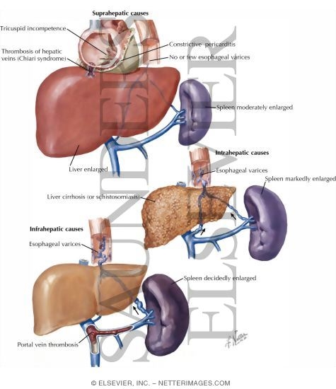
a) Pre-hepatic Causes :
Portal vein thrombosis - seen in umbilical sepsis (infants)
Splenic vein thrombosis - Complication of pancreatitis, pancreatic tumour
b) Intrahepatic Causes :
i) Pre-sinusoidal :
Schistosomiasis
Primary biliary cirrhosis
Chronic active hepatitis
Sarcoidosis
ii) Sinusoidal :
Cirrhosis
Cytotoxic drugs
Vitamin A intoxication
iii) Post-sinusoidal :
Cirrhosis
Veno-occlusive diseases
c) Post-hepatic causes :
Budd-Chiari's syndrome
Tricuspid regurgitation
Constrictive Pericarditis
Clinical presentation (History and Examination)
Malnutrition
Ascites
Hematemesis and Malena
Encephalopathy
Caput medusae
Splenomegaly
Venous hum heard
Look for signs and symptoms of chronic liver disease
How do you manage these patients?
1) Esophageal varices without prior h/o of bleeding
Medical management is ideal in such cases.
Start Propanolol orally to reduce portal pressure, provided that there's no contraindication against B-blockers.
If contraindication present, isosorbide-5-mononitrate is an alternative.
Studies have shown that B-blockers reduces 45% of the risk of bleeding.
2) Ruptured esophageal varices presented with hematemesis
95% of the cases - originating from the esophageal varices, 5% - gastric origin
First, assess the rate and volume of bleeding :
Take pulse and BP in standing and sitting position
Gain IV ascess - Blood is withdrawn for hematocrit, coagulation profile, LFT and BUSE, blood grouping and cross matching
Provide immediate fluid resuscitation (Crystalloids, colloids, or even blood transfusion)
Insert CVP line - for ease of rapid transfusion later to prevent volume overload
Start Vasopressin IV (Contraindicated in angina) or Somastostatin IV.
Or Octreotide IV (more potent and duration of action is longer)
Usually 3 days later, as the patient's condition has stabilised, start B-blocker to reduce portal pressure and prevent further bleeding.
Plan for endoscopic treatment :
a) Band ligation
b) Sclerotherapy (Sodium Tetradecyl Sulphate - STS)
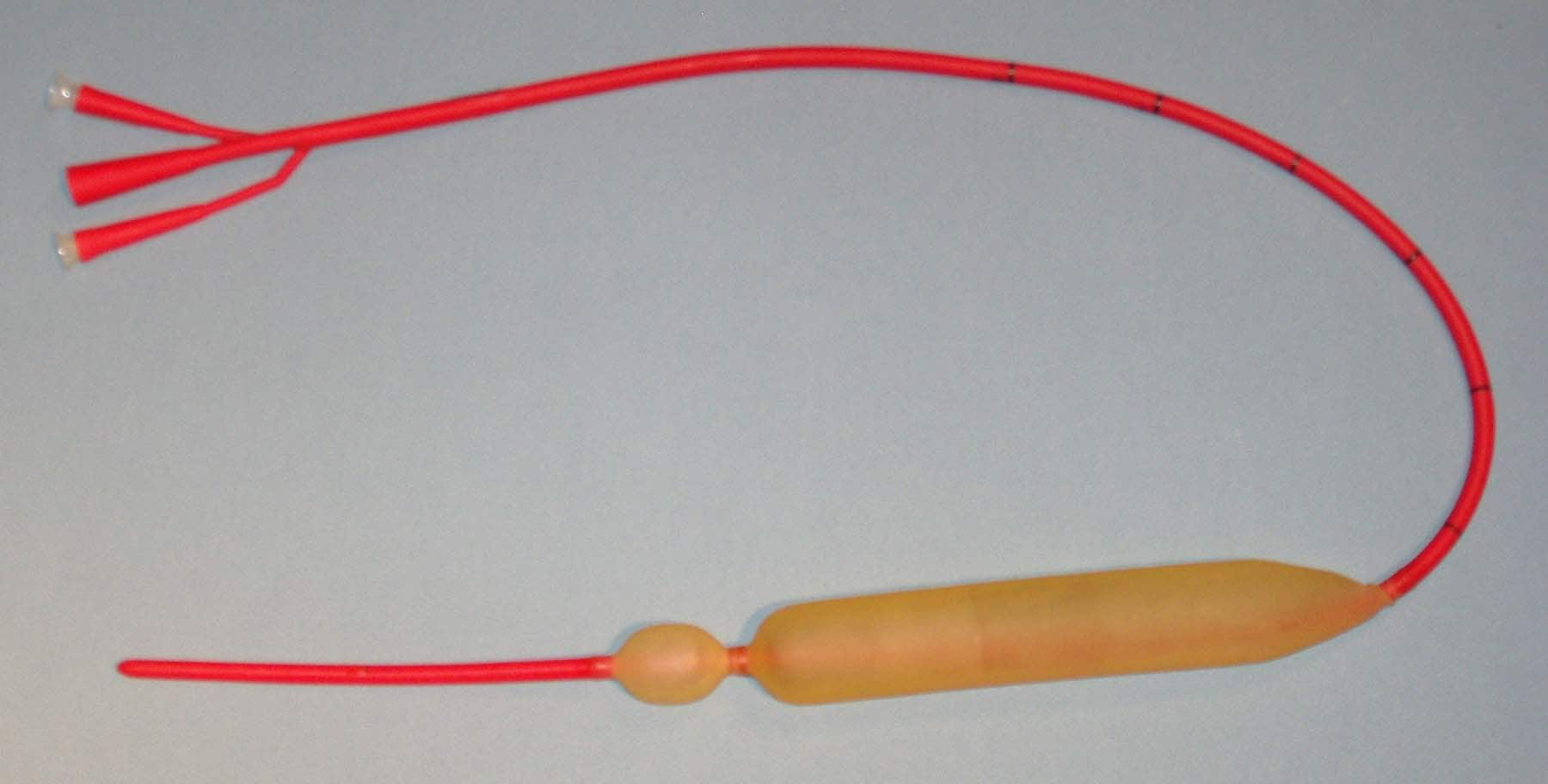
If the patient is not reponsive to the above measures and still bleeding or endoscopic intervention is not available (district hospitals), a Sangstaken-Blakemore tube can be inserted to prevent bleeding to buy time for deciding what's the next step. (Shoiuld be removed after 48 hrs)
Start oral neomycin (to reduce bowel flora -> less conversion of nitrogenous waste within bowel back to ammonia -> prevent hyperammonemia)
Start lactulose (to reduce bowel transit time)
Repeat every 2 weeks the sclerotherapy/band ligation until all the varices have been treated.
Monday, November 23, 2009
Sunday, November 22, 2009
Rectal bleeding
Rectal bleeding usually indicates lower GI bleeding (below duodenojejunal junction). Bear in mind that any patient, aged >45 yrs old, with complaints of :
colickly abdominal pain, PR bleeding and changes in bowel habits
Colorectal CA must be considered unless proven otherwise.
Some causes of rectal bleeding
1) Anal cause
a) Haemorrhoids
Piles are very common.
Uncomplicated piles are not painful.
b) Fissure-in-ano
Fissure-in-ano usually causes painful defecation.
The precipitating cause is usually constipation, hence constipation is usually worsened by the patient fear of passing motion, since it's painful.
Pain usually persists for minutes or even hours after defecation.
c) Carcinoma
The history of anal carcinoma is similar to that of Fissure-in-ano.
However, it's usually seen in the elderly.
d) Trauma
History of penetrating injury into the anus.
Sexual abuse? Anal intercourse?
2) Colorectal causes
a) Carcinoma
As mentioned in previous posts
b) Polyps
The history given by patient is usually the same as carcinoma
c) Diverticulitis
The difference between Diverticulitis and Carcinoma :
Duration : Diverticulitis = longer, Carcinoma = shorter
Pain : Diverticulitis = usually painful, Carcinoma = painless (initial)
Bleeding pattern : Diverticulitis = periodic, massive
Carcinoma = usually smaller in amount, persistent
Mass per abdomen : Diverticulitis = tender, Carcinoma = tenderless
Abdominal radiograph : Diverticulitis = diffuse changes, Carcinoma = localised
d) Inflammatory bowel disease
Usually presented as sudden onset of watery diarrhoea, together with brown stools, mucous, and blood. Ulcerative proctitis can presents as tenesmus
e) Ischaemic colitis, angiodysplasia
f) Irradiative colitis
Especially common in patients with pelvic malignancies, due to irradiation
g) Rectal prolapse
Patient usually complaints of something hanging out at his/her back opening besides PR bleeding
3) Small bowel
a) Meckel's diverticulum
Consider this diagnosis in young adults with frequent painless PR bleeding
b) Acute mesenteric infarction
Patient with h/o of cardiac disease (embolism), with complaints of diffuse abdominal pain, PR bleeding, collapse, with signs of shock, etc -> consider this as well
4) Massive Upper GI bleeding
Due to the massive nature of the upper GI bleeding, the intestinal transit is fast, and hence instead of presenting with hematemesis, patient presents with massive PR bleeding, with shock-like features
5) Bleeding disorders
6) Drugs (anticoagulants)
7) Uremic bleeding
8) Infective causes - dysentry
colickly abdominal pain, PR bleeding and changes in bowel habits
Colorectal CA must be considered unless proven otherwise.
Some causes of rectal bleeding
1) Anal cause
a) Haemorrhoids
Piles are very common.
Uncomplicated piles are not painful.
b) Fissure-in-ano
Fissure-in-ano usually causes painful defecation.
The precipitating cause is usually constipation, hence constipation is usually worsened by the patient fear of passing motion, since it's painful.
Pain usually persists for minutes or even hours after defecation.
c) Carcinoma
The history of anal carcinoma is similar to that of Fissure-in-ano.
However, it's usually seen in the elderly.
d) Trauma
History of penetrating injury into the anus.
Sexual abuse? Anal intercourse?
2) Colorectal causes
a) Carcinoma
As mentioned in previous posts
b) Polyps
The history given by patient is usually the same as carcinoma
c) Diverticulitis
The difference between Diverticulitis and Carcinoma :
Duration : Diverticulitis = longer, Carcinoma = shorter
Pain : Diverticulitis = usually painful, Carcinoma = painless (initial)
Bleeding pattern : Diverticulitis = periodic, massive
Carcinoma = usually smaller in amount, persistent
Mass per abdomen : Diverticulitis = tender, Carcinoma = tenderless
Abdominal radiograph : Diverticulitis = diffuse changes, Carcinoma = localised
d) Inflammatory bowel disease
Usually presented as sudden onset of watery diarrhoea, together with brown stools, mucous, and blood. Ulcerative proctitis can presents as tenesmus
e) Ischaemic colitis, angiodysplasia
f) Irradiative colitis
Especially common in patients with pelvic malignancies, due to irradiation
g) Rectal prolapse
Patient usually complaints of something hanging out at his/her back opening besides PR bleeding
3) Small bowel
a) Meckel's diverticulum
Consider this diagnosis in young adults with frequent painless PR bleeding
b) Acute mesenteric infarction
Patient with h/o of cardiac disease (embolism), with complaints of diffuse abdominal pain, PR bleeding, collapse, with signs of shock, etc -> consider this as well
4) Massive Upper GI bleeding
Due to the massive nature of the upper GI bleeding, the intestinal transit is fast, and hence instead of presenting with hematemesis, patient presents with massive PR bleeding, with shock-like features
5) Bleeding disorders
6) Drugs (anticoagulants)
7) Uremic bleeding
8) Infective causes - dysentry
An approach to a case of Hematemesis
Hematemesis means vomiting of blood.
It can be either a frank blood, or altered, coffee-ground coloured blood (altered by digestive enzyme)
The aetiology of hematemesis is usually proximal to the duodenojejunal junction.
Causes
1) Swallowed blood
Due to hemoptysis, epitaxis
2) Oesophageal causes
Ruptured oesophageal varices
Reflux oesophagitis
Esophageal carcinoma
3) Stomach and duodenum
Peptic ulcer disease
Mallory-Weiss disease
Acute gastric erosions
Gastric carcinoma
4) Bleeding disorders
Hemophilia
Thrombocytopoenia
Coagulopathy (due to liver disease)
5) Drugs
NSAIDS
Aspirin
Steroids
Anticoagulants
6) Others
Uremia
Connective tissue disorders
History : Important questions
1) Is there any h/o of epitaxis, hemoptysis?
2) Ask for h/o of chronic liver disease
3) Any retrosternal burning chest pain radiating upwards or heartburn?
4) Any h/o of dysphagia, odynophagia, weight lost?
5) Ask for h/o of peptic ulcer disease
6) Any h/o of consumption of large meal and alcohol?
7) Does the hematemesis preceded by severe bouts of vomiting?
8) Ask for h/o of anaemia
9) Any recent h/o of acute pancreatitis? Any head injuries? (Cushing's ulcer) Or Any h/o of burns? (Curling's ulcer)
10) Any h/o of bleeding disorders? In the family, is there any?
11) Any h/o of drug intake?
12) Ask for symptoms of uraemia
On examination
1) Depends on the severity of bleeding, does the patients appears to be in shock?
Cool extremities
Prolonged capillary filling time
Tachycardia
Hypotension
Reduced skin turgosity
Altered sensorium
Sunken eyeballs
Dry tongue
Reduced urine output
2) Check around the nose - is there any blood?
3) Examine the chest for any cause of hemoptysis
4) Look for pallor
5) Look for signs of chronic liver disease
6) Any epigastric mass, palpable Left SC nodes?
7) Any epigastric tenderness?
8) Any bruises? Any signs of uremia?
Investigations
1) Full blood count, ESR
Hb level, platelet count, any raised ESR? (connective tissue disorders)
2) Liver function test
3) Coagulation profile (PT and INR)
4) BUSE (Renal profile)
5) Oesophagogastroduodenoscopy (OGD)
It can be either a frank blood, or altered, coffee-ground coloured blood (altered by digestive enzyme)
The aetiology of hematemesis is usually proximal to the duodenojejunal junction.
Causes
1) Swallowed blood
Due to hemoptysis, epitaxis
2) Oesophageal causes
Ruptured oesophageal varices
Reflux oesophagitis
Esophageal carcinoma
3) Stomach and duodenum
Peptic ulcer disease
Mallory-Weiss disease
Acute gastric erosions
Gastric carcinoma
4) Bleeding disorders
Hemophilia
Thrombocytopoenia
Coagulopathy (due to liver disease)
5) Drugs
NSAIDS
Aspirin
Steroids
Anticoagulants
6) Others
Uremia
Connective tissue disorders
History : Important questions
1) Is there any h/o of epitaxis, hemoptysis?
2) Ask for h/o of chronic liver disease
3) Any retrosternal burning chest pain radiating upwards or heartburn?
4) Any h/o of dysphagia, odynophagia, weight lost?
5) Ask for h/o of peptic ulcer disease
6) Any h/o of consumption of large meal and alcohol?
7) Does the hematemesis preceded by severe bouts of vomiting?
8) Ask for h/o of anaemia
9) Any recent h/o of acute pancreatitis? Any head injuries? (Cushing's ulcer) Or Any h/o of burns? (Curling's ulcer)
10) Any h/o of bleeding disorders? In the family, is there any?
11) Any h/o of drug intake?
12) Ask for symptoms of uraemia
On examination
1) Depends on the severity of bleeding, does the patients appears to be in shock?
Cool extremities
Prolonged capillary filling time
Tachycardia
Hypotension
Reduced skin turgosity
Altered sensorium
Sunken eyeballs
Dry tongue
Reduced urine output
2) Check around the nose - is there any blood?
3) Examine the chest for any cause of hemoptysis
4) Look for pallor
5) Look for signs of chronic liver disease
6) Any epigastric mass, palpable Left SC nodes?
7) Any epigastric tenderness?
8) Any bruises? Any signs of uremia?
Investigations
1) Full blood count, ESR
Hb level, platelet count, any raised ESR? (connective tissue disorders)
2) Liver function test
3) Coagulation profile (PT and INR)
4) BUSE (Renal profile)
5) Oesophagogastroduodenoscopy (OGD)
Saturday, November 21, 2009
Short cases - Lumps
1) Dermoid cyst
Dermoid cyst is a cyst located deep to the skin and lined by the skin.
It forms either due to accident during antenatal development or even following injury, some skin is being implanted into the subcutaneous tissue.
Hence, dermoid cyst can be congenital or accquired.
a) Congenital dermoid cyst
History
May be noticed at birth, or years later when it gradually distends to a noticable size. The common complaints, usually by the parents is cosmetic disfigurement since it's a swelling at the neck and face. Rarely it becomes large enough to cause mechanical disability or affecting the vision.
On examination
Congenital dermoid cyst is usually formed when the skin dermatome fuses.
It's commonly found at the midline trunk, face and neck, outer or inner aspect of eye brow, or behind the ear.
Shape is usually spherical, with diameter of 1-2cm.
Smooth surfaced.
Congenital dermoid cyst over the face is usually soft.
Since it usually doesn't contain clear fluid as it supposed to be (mixture of sebum, sweat and desquamated epithelial cells), it doesn't transilluminate.
It fluctuates and if it's large enough, there's fluid thrill as well.
Skin over the cyst is pinchable.
Non-pulsatile, non-compressible, non-reducible.
Local lymph nodes are not enlarged.
b) Accquired implantational dermoid cyst
History
There's history of old injury, such as deep cut, stab injuries and etc.
These implantational dermoid cyst is usually found over areas susceptible to repeated traumas, eg the fingers.
Hence, it can be painful, or even interferes with gripping and touch.
Examination
Seen over sites liable to repeated trauma - beneath skin of fingers.
Size and shape - Spherical, small with diameter 0.5-1cm
Smoothed surace
Due to it's small size, it's almost impossible to elicit specific signs for cystic swelling, which is fluctuation and fluid thrill.
It's usually hard, and the skin overlying it is usually scarred.
The skin is either tethered deep to the scar or within it.
It's mobile over the deeper structures, which is usually normal.
Commonly confused with sebaceous cyst, but with an old scar and h/o of injury is significant for diagnosis.
2) Subcutaneous Abscess

Dermoid cyst is a cyst located deep to the skin and lined by the skin.
It forms either due to accident during antenatal development or even following injury, some skin is being implanted into the subcutaneous tissue.
Hence, dermoid cyst can be congenital or accquired.
a) Congenital dermoid cyst
History
May be noticed at birth, or years later when it gradually distends to a noticable size. The common complaints, usually by the parents is cosmetic disfigurement since it's a swelling at the neck and face. Rarely it becomes large enough to cause mechanical disability or affecting the vision.
On examination
Congenital dermoid cyst is usually formed when the skin dermatome fuses.
It's commonly found at the midline trunk, face and neck, outer or inner aspect of eye brow, or behind the ear.
Shape is usually spherical, with diameter of 1-2cm.
Smooth surfaced.
Congenital dermoid cyst over the face is usually soft.
Since it usually doesn't contain clear fluid as it supposed to be (mixture of sebum, sweat and desquamated epithelial cells), it doesn't transilluminate.
It fluctuates and if it's large enough, there's fluid thrill as well.
Skin over the cyst is pinchable.
Non-pulsatile, non-compressible, non-reducible.
Local lymph nodes are not enlarged.
b) Accquired implantational dermoid cyst
History
There's history of old injury, such as deep cut, stab injuries and etc.
These implantational dermoid cyst is usually found over areas susceptible to repeated traumas, eg the fingers.
Hence, it can be painful, or even interferes with gripping and touch.
Examination
Seen over sites liable to repeated trauma - beneath skin of fingers.
Size and shape - Spherical, small with diameter 0.5-1cm
Smoothed surace
Due to it's small size, it's almost impossible to elicit specific signs for cystic swelling, which is fluctuation and fluid thrill.
It's usually hard, and the skin overlying it is usually scarred.
The skin is either tethered deep to the scar or within it.
It's mobile over the deeper structures, which is usually normal.
Commonly confused with sebaceous cyst, but with an old scar and h/o of injury is significant for diagnosis.
2) Subcutaneous Abscess
History
Throbbing pain which steadily worsens, and keeps patient awaken at night.
Patient usually notice a swelling at the site of pain.
May complaints of fever with chills and rigor.
It may rupture and discharging pus out of the skin before they seek medical attention.
The patient may have h/o of diabetes, having debilitating diseases, or even IV drug use.
Examination
Since the buttocks and upper thigh are usual sites of injection, abscess may be formed there. And in IV drug users, over cubital fossa or groin.
Skin over swelling appears red and shinny.
Surface is not definate.
Usually started as a patch of induration, which later as pus collects, a spherical mass is formed.
There's local rise in temperature.
The edge is not palpable since due to the induration and the edema usually fuses with the normal tissue.
It's tender.
Initially it feels hard, when pus started to collect, it becomes soft at the centre and fluctuates.
Skin over swelling is not pinchable.
Regional lymph nodes may be enlarged and tender
3) Sebaceous cyst
Our skin is kept oily and soft by secretions of sebum from sebaceous gland.
The mouth of the sebaceous glands are located at the hair follicles.
Any blockage over these mouth can result in it's distension within it's own secretion and results in formation of sebaceous cyst.
History
Rarely present before adolescence, since it's a slow-growing swelling.
Usually seen in young adults or middle-aged individual.
It's usually detected incidentally by patients as they're combing their hair, when they complained of scratched lump.
It can be infected, where the size suddenly increased rapidly.
Sebum secreted from wide punctum can be later hardened to form a sebaceous horn.
Examination
It's usually present at scalp, back, shoulders, and scrotum. (Never in the palm and soles since there's no sebaceous gland over these areas)
Skin over swelling is normal unless infected.
Shape - spherical, with smooth surface.
Temperature is not raised and is not tender unless it gets infected.
Edge is easily felt, well-defined.
Consistency - hard.
No fluctuations or fluid thrill.
As the swelling increases in size, the point of fixation will be drawn inwards and punctum is formed. Punctum is diagnostic for sebaceous cyst, but however, only one-half of such swelling presents with a punctum.
Skin over swelling is not pinchable.
Local lymph nodes are not palpable.
4) Lipoma
History
Lipoma is a slow-growing swelling, rarely regresses.
Occurs at any age, but relatively uncommon in children.
Not associated with any symptoms, but presents to the doctor usually because they have noticed a lump and wanted to know what it is.
Patients can have multiple lipoma (lipomatosis), usually over the neck and buttocks.
Examination
Common sites of lipoma includes the upper and lower limb, back, buttocks, neck, etc.
Size is variable, shape - hemiovoid, spherical, etc.
On inspection, surface appears smooth. But when the swelling is palpated carefully, especially when firm pressure is applied, it's lobulated and depression in between these lobulations is seen.
Edge is soft, compressible and tends to slip away from examining hands (slip sign)
Composition - solid fat (fat in body temperature is solid instead of fluid)
Consistency - soft
There's pseudofluctuation, since it's consistency is soft. However, one will notice that on gentle pressure,the plane of swelling over palpating fingers are not tense or not buldging out.
There may be pseudo-transillumination.
Skin over swelling is pinchable.
As muscle is tensed, it may either be more prominent or less (it can arise above or beneath the muscle)
Compressible swelling.
No enlargement of regional lymph nodes.
History taking and examination of a swelling
Some key questions to be asked regarding a swelling (generally)
1. When do you first notice the lump?
REMEMBER, first noticed the lump 3 months ago is not the same as first appeared 3 months ago.
2. How do you notice it?
Below are the 3 commonest answers :
a) It's painful
b) I noticed it accidentally
c) Others told me about it
Generally, if the lump is painful, the commonest aetiology is inflammation.
Most of the patients thought that only painful lumps are cancerous.
3. How does the lump disturbs you?
Basically, the question is asking about the associated symptoms.
It can be pain, discharge, dysphagia, dyspnoea, cosmetically disfiguring, fear of malignancy, etc.
4. Any changes to the lump since you first notice it?
The commonest change is the size.
Whether the lump has increased or decreased in size, or it's size fluctuates.
5. Has the lump ever dissapears before?
Does the lump dissapears when the patient is lying down supine?
or any other activities
6. Do you ever had any other lumps before this?
Asking for multiplicity
7. What do you think is the cause?
Particularly important if there's history of trauma
On examination :
Note the -
a) Position
b) Colour and texture of skin over swelling
c) Size
d) Shape
e) Surface
f) Temperature
g) Tenderness
h) Edge - indistinct/well-defined
i) Composition
Calcified tissues/bone makes the swelling hard
Swelling packed with cells : Firm
Or it might contain fluid (lymph, blood, pus)
Intravascular blood
Gas
j) Consistency
Stony hard - not indentable at all, as hard as bone
Firm - hard, but not as hard as bone
Rubbery - slightly indentable, feels like rubber
Spongy - indentable, but with some resillence
Soft - Squashable, no resillence
k) Fluctuation
Palpate the swelling over 3 planes.
Pressure over the 3rd plane of the swelling usually causes the other 2 planes to buldge out or tensed-up
Positive fluctuation test indicates that the swelling might contains fluid
l) Fluid thrill
Only swelling which contains fluid transmits percussion waves.
Use one of the finger to tap one end of the lump, and feel for the vibration produced at another end, using another finger from another hand.
If the swelling is too large, the percussion wave might be transmitted through the wall.
Hence, one should place a hand at the middle of the swelling to prevent such transmission.
m) Transillumination
If the swelling contains clear fluid, it transilluminate.
It should be done using a small bright light source, in a dark room.
Eg, hydrocele, epididymal cyst
n) Pulsatility
Place one finger of each hand over two ends of the swelling.
If both the fingers are moving upwards and outwards -> expansile (eg, aneurysm)
If both the fingers are moving only upwards (one direction) -> transmitted (a lump overlying an artery)
o) Compressibility
As pressure is applied to the swelling, it's compressed.
But once the pressuring hand is removed, the swelling immediately reappears.
p) Bruits
q) Reducibility
This is different from compressibility.
If the swelling is reducible, it is reduced into another space.
As the pressuring hand is removed, the swelling usually takes some time before reappearing, or will only reappear when there's stimulus, eg cough
r) Relation with surrounding tissue
Is it pinchable from the skin?
When the muscle is tensed,
Does it becomes more prominent, less mobile or less prominent and less easily felt?
When the swelling overlies a nerve/or artery -> not mobile along it's course, but mobile across it's length
s) Any palpable regional lymph nodes?
t) General examination
1. When do you first notice the lump?
REMEMBER, first noticed the lump 3 months ago is not the same as first appeared 3 months ago.
2. How do you notice it?
Below are the 3 commonest answers :
a) It's painful
b) I noticed it accidentally
c) Others told me about it
Generally, if the lump is painful, the commonest aetiology is inflammation.
Most of the patients thought that only painful lumps are cancerous.
3. How does the lump disturbs you?
Basically, the question is asking about the associated symptoms.
It can be pain, discharge, dysphagia, dyspnoea, cosmetically disfiguring, fear of malignancy, etc.
4. Any changes to the lump since you first notice it?
The commonest change is the size.
Whether the lump has increased or decreased in size, or it's size fluctuates.
5. Has the lump ever dissapears before?
Does the lump dissapears when the patient is lying down supine?
or any other activities
6. Do you ever had any other lumps before this?
Asking for multiplicity
7. What do you think is the cause?
Particularly important if there's history of trauma
On examination :
Note the -
a) Position
b) Colour and texture of skin over swelling
c) Size
d) Shape
e) Surface
f) Temperature
g) Tenderness
h) Edge - indistinct/well-defined
i) Composition
Calcified tissues/bone makes the swelling hard
Swelling packed with cells : Firm
Or it might contain fluid (lymph, blood, pus)
Intravascular blood
Gas
j) Consistency
Stony hard - not indentable at all, as hard as bone
Firm - hard, but not as hard as bone
Rubbery - slightly indentable, feels like rubber
Spongy - indentable, but with some resillence
Soft - Squashable, no resillence
k) Fluctuation
Palpate the swelling over 3 planes.
Pressure over the 3rd plane of the swelling usually causes the other 2 planes to buldge out or tensed-up
Positive fluctuation test indicates that the swelling might contains fluid
l) Fluid thrill
Only swelling which contains fluid transmits percussion waves.
Use one of the finger to tap one end of the lump, and feel for the vibration produced at another end, using another finger from another hand.
If the swelling is too large, the percussion wave might be transmitted through the wall.
Hence, one should place a hand at the middle of the swelling to prevent such transmission.
m) Transillumination
If the swelling contains clear fluid, it transilluminate.
It should be done using a small bright light source, in a dark room.
Eg, hydrocele, epididymal cyst
n) Pulsatility
Place one finger of each hand over two ends of the swelling.
If both the fingers are moving upwards and outwards -> expansile (eg, aneurysm)
If both the fingers are moving only upwards (one direction) -> transmitted (a lump overlying an artery)
o) Compressibility
As pressure is applied to the swelling, it's compressed.
But once the pressuring hand is removed, the swelling immediately reappears.
p) Bruits
q) Reducibility
This is different from compressibility.
If the swelling is reducible, it is reduced into another space.
As the pressuring hand is removed, the swelling usually takes some time before reappearing, or will only reappear when there's stimulus, eg cough
r) Relation with surrounding tissue
Is it pinchable from the skin?
When the muscle is tensed,
Does it becomes more prominent, less mobile or less prominent and less easily felt?
When the swelling overlies a nerve/or artery -> not mobile along it's course, but mobile across it's length
s) Any palpable regional lymph nodes?
t) General examination
Urology - Q & A
What are the common causes of urinary tract obstruction?
Upper UT obstruction :
Renal, ureteric calculi
Pelvi-ureteric junction obstruction
Retroperitoneal fibrosis (idiopathic, malignancy)
Transitional cell carcinoma
Congenital (Ectopic ureter, ureterocele)
Infections (Schistosomiasis, TB)
Lower UT obstruction :
Urethral causes (stricture, tumour, stone)
Prostate (BPH, CA prostate)
Bladder neck (CA, stones, neurological causes, stricture)
What are the clinical features of acute urinary retention?
Patient usually complains of suprapubic pain, unable to pass urine
Well aware that there's bladder distension
On examination, bladder is palpable with the following features :
An ovoid mass originating from the pelvis
Can't get below the swelling
Tense, tender, smooth surface
Gentle pressure increases desire to micturate
Immobile
Percussion - dull
Positive fluid thrill
Sometimes, the bladder can be extending up to the umbilicus
(in cases of acute on chronic urinary retention)
What are the clinical features of chronic urinary retention?
Bear in mind that there're 2 types of chronic UR :
a) High pressure type
Associated with chronic bladder outflow obstruction
Can progress to hydroureter and hydronephrosis and eventually, obstructive renal failure
b) Low pressure type
Associated with bladder atony
Doesn't progress towards renal failure
Symptom-wise, patient is usually unaware that there's bladder distension
Usually, such condition is painless
However, there's both obstructive and irritative symptoms of micturition (as in BPH)
And, there's overflow incontinence :
The patient can pass an apparently normal volume of urine
However, whenever there's raised intra-abdominal pressure, there's dribbling of urine
On examination :
Bladder is usually palpable, upto the umbilicus
Non-tender, not tense
Gentle pressure may not be associated with any increased desire in micturition
Dull note on percussion, +ve fluid thrill
Briefly discuss about the clinical significance of urodynamic studies
Upper UT obstruction :
Renal, ureteric calculi
Pelvi-ureteric junction obstruction
Retroperitoneal fibrosis (idiopathic, malignancy)
Transitional cell carcinoma
Congenital (Ectopic ureter, ureterocele)
Infections (Schistosomiasis, TB)
Lower UT obstruction :
Urethral causes (stricture, tumour, stone)
Prostate (BPH, CA prostate)
Bladder neck (CA, stones, neurological causes, stricture)
What are the clinical features of acute urinary retention?
Patient usually complains of suprapubic pain, unable to pass urine
Well aware that there's bladder distension
On examination, bladder is palpable with the following features :
An ovoid mass originating from the pelvis
Can't get below the swelling
Tense, tender, smooth surface
Gentle pressure increases desire to micturate
Immobile
Percussion - dull
Positive fluid thrill
Sometimes, the bladder can be extending up to the umbilicus
(in cases of acute on chronic urinary retention)
What are the clinical features of chronic urinary retention?
Bear in mind that there're 2 types of chronic UR :
a) High pressure type
Associated with chronic bladder outflow obstruction
Can progress to hydroureter and hydronephrosis and eventually, obstructive renal failure
b) Low pressure type
Associated with bladder atony
Doesn't progress towards renal failure
Symptom-wise, patient is usually unaware that there's bladder distension
Usually, such condition is painless
However, there's both obstructive and irritative symptoms of micturition (as in BPH)
And, there's overflow incontinence :
The patient can pass an apparently normal volume of urine
However, whenever there's raised intra-abdominal pressure, there's dribbling of urine
On examination :
Bladder is usually palpable, upto the umbilicus
Non-tender, not tense
Gentle pressure may not be associated with any increased desire in micturition
Dull note on percussion, +ve fluid thrill
Briefly discuss about the clinical significance of urodynamic studies
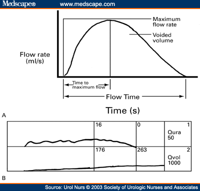
The maximum urinary flow rate can be determined by urodynamic studies.
In men, it's 15-30 ml/sec; in females, it's 20-40 ml/sec.
A graph of urinary flow rate versus time can be plotted, in which different pattern of curves usually indicates different causes of obstruction.
A normal urinary flow : Rises to the peak rapidly, and rapidly drops down to the baseline (as shown above)
In bladder outflow obstruction, there's prolonged rise to poor maximum flow rate and periods of prolonged variability in flow rate
In urethral obstruction, there's a stable, plateu-shaped curve, with prolonged flow rate.
Together with cystometry, the bladder capacity, capacity during desire to micturate, pressure on the detrusor muscle in full bladder, residual urine volume can be determined.
History taking in a case of hematuria
First of all, the causes of hematuria :
1) Kidney
Glomerular diseases
Polycystic kidney disease
Kidney stones
Trauma (renal biopsy)
Renal adenocarcinoma
Renal TB
Renal vein thrombosis
Embolism
2) Ureter
Ureteric stones
Neoplasm
3) Bladder
Bladder stones
CA bladder
Bladder Trauma
Inflammation (Cystitis, stones, TB)
4) Prostate
Benign Prostatic Hyperplasia
CA prostate
5) Urethra
Urethral trauma
Urethral stones
Neoplasm
Trauma
General causes
Anticoagulants (Warfarin)
Thrombocytopoenia
Sickle cell disease
Malaria
Schistosomiasis
Blood dyscarias (Hemophilia)
Sternous exercises
Red urine
Hemoglobinuria
Myoglobinuria
Beetroot
Senna
Rifampicin
Phenopthalein
History : Important questions!
1) Is there any pain on micturition?
2) Is it :
Painless hematuria? (Renal TB, RCC, CA bladder)
Total hematuria? (suggestive of bleeding from upper urinary tract)
Initial hematuria? (bleeding from lower urinary tract = prostate?urethra?)
Terminal hematuria? (bleeding from prostate?bladder?)
3) Is there family history of renal disease? (Polycystic)
4) Is there any h/o of drug intake? (anticoagulant)
5) Is there any h/o of substance ingestion that might cause red discolouration of urine?
6) Is there travel history? (Malaria/Schistosomiasis)
7) Is there any fixed loin pain? (Renal causes)
8) Is there any colicky loin to groin pain? (Ureteric colic)
9) Any symptoms suggestive of bladder stones? (Frequency, suprapubic pain, etc)
10) Any symptoms of prostatism?
11) Any h/o of trauma? (including renal biopsy)
12) Any h/o of general debility? (malaise, lost of weight, appetite, etc)
13) Any h/o of bleeding disorders?
14) Any h/o of TB in other parts of body? (esp. pulmonary)
15) Any h/o of sternous exercises done recently?
16) Any h/o of dehydration?
Investigations
1) Full blood count
2) ESR
3) Urine microscopy
4) Chest X-ray
5) Coagulation profile (PT and INR)
6) KUB film or IVU (calculus)
1) Kidney
Glomerular diseases
Polycystic kidney disease
Kidney stones
Trauma (renal biopsy)
Renal adenocarcinoma
Renal TB
Renal vein thrombosis
Embolism
2) Ureter
Ureteric stones
Neoplasm
3) Bladder
Bladder stones
CA bladder
Bladder Trauma
Inflammation (Cystitis, stones, TB)
4) Prostate
Benign Prostatic Hyperplasia
CA prostate
5) Urethra
Urethral trauma
Urethral stones
Neoplasm
Trauma
General causes
Anticoagulants (Warfarin)
Thrombocytopoenia
Sickle cell disease
Malaria
Schistosomiasis
Blood dyscarias (Hemophilia)
Sternous exercises
Red urine
Hemoglobinuria
Myoglobinuria
Beetroot
Senna
Rifampicin
Phenopthalein
History : Important questions!
1) Is there any pain on micturition?
2) Is it :
Painless hematuria? (Renal TB, RCC, CA bladder)
Total hematuria? (suggestive of bleeding from upper urinary tract)
Initial hematuria? (bleeding from lower urinary tract = prostate?urethra?)
Terminal hematuria? (bleeding from prostate?bladder?)
3) Is there family history of renal disease? (Polycystic)
4) Is there any h/o of drug intake? (anticoagulant)
5) Is there any h/o of substance ingestion that might cause red discolouration of urine?
6) Is there travel history? (Malaria/Schistosomiasis)
7) Is there any fixed loin pain? (Renal causes)
8) Is there any colicky loin to groin pain? (Ureteric colic)
9) Any symptoms suggestive of bladder stones? (Frequency, suprapubic pain, etc)
10) Any symptoms of prostatism?
11) Any h/o of trauma? (including renal biopsy)
12) Any h/o of general debility? (malaise, lost of weight, appetite, etc)
13) Any h/o of bleeding disorders?
14) Any h/o of TB in other parts of body? (esp. pulmonary)
15) Any h/o of sternous exercises done recently?
16) Any h/o of dehydration?
Investigations
1) Full blood count
2) ESR
3) Urine microscopy
4) Chest X-ray
5) Coagulation profile (PT and INR)
6) KUB film or IVU (calculus)
Subscribe to:
Posts (Atom)