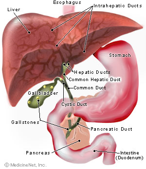
Pathophysiology
Mostly cholesterol stones (80%), pigment stones or mixed stones.
Whether cholesterol remains as solution within bile depends on it's concentration, and the levels of phospholipids and bile acids within.
If the bile is supersaturated by cholesterol, and/or levels of phospholipids and bile acids is low, this promotes formation of cholesterol crystals.
These cholesterol crystals are toxic towards the gall bladder musculature, and hence damages it.
This results in gall bladder hypomotility, and enhances the nucleation of theses gall bladder crystals.
Eventually, gall stones are formed.
As for pigment stones, there are 2 types, namely black and brown stones.
Black stones are sterile stones, usually caused by extensive hemolysis leading to unconjugated hyperbilirubinemia. Eg, seen in Hereditary Spherocytosis, Sickle-cell disease.
Whilst brown stones are non-sterile stones, mainly caused by infection.
Eg, certain bacteria (E.coli) produces B-glucoronidase, which converts conjugated bilirubin back to unconjugated bilirubin.
Also associated with parasitic worm infestation, eg : Ascariasis, Clonorchis sinensis
Complications
1) Gall bladder
Biliary colic
Acute cholecystitis
Chronic cholecystitis
Empyema
Mucocele
Perforation
2) Biliary tract
Biliary tract obstruction
Acute pancreatitis
Ascending cholangitis
3) Intestine
Gall stone ileus
Acute cholecystitis
Usually caused by obstruction of cystic duct by gall stones, leading to gall bladder distension, chemical inflammation, and eventual bacterial infection.
History
Age : Typically 30-60 years old. Presentation in younger patients, may be due to Congenital hemolytic anemia
Gender : Females are more commonly affected
Symptoms :
Fever
Sudden onset of severe, continuous RHC pain
Radiates to the back (close to the inferior angle of right scapula)
Associated with nausea and vomiting
Duration of pain usually exceeds 3-6 hours
Pain aggravated by movements and breathing
May have previous h/o of flatulent dyspepsia or biliary colic
On general examination :
Patient appears ill
Lying still on bed, breathing shallowly
Tachycardia +ve, Pyrexia +ve
During initial stages of inflammation - RHC fullness (known as Zackary-cope's sign)
RHC tenderness, guarding/rigidity, +ve Murphy's sign
Before the onset of guarding, during the early stages the gall bladder may be palpable
If the inflammation persisted for a few days, with subsequent subside of symptoms, an inflammatory mass may be palpable (empyema)
Boas Sign +ve
Biliary colic
Before proceeding to investigations of Acute cholecystitis, briefly about biliary colic.
It's actually a misnomer, caused by spasm of gall bladder musculature, trying to force the stone down the cystic duct.
Since it's a visceral pain, pain is usually felt over the epigastrium (foregut)
The pain is typically aggravated by intake of oily food.
Since after ingesting oily food, as it passes through the 2nd part of duodenum, it stimulates the production of cholecystikinin from the duodenal mucosa.
It causes contraction of the gall bladder musculature over the stones, hence causing pain.
Duration of pain typically lasted < 3 hours.
However, in practice, patient usually localizes their pain poorly (c/c usually diffuse upper abdominal pain)
It's a pseudocolic, since there's no complete relief of pain in between periods of excruciating exacerbations.
There might be nausea and vomiting.
On examination, there may be tachycardia, but fever is absent.
On abdominal examination, other than tenderness, guarding, usually there's no other signs.
Hence, there's a frequent overlap in between the clinical features of biliary colic and acute cholecystitis.
Here the table showing their difference :
Investigations
Full blood count - reveals leucocytosis
Abdominal X ray - not useful, only 10-15% of the calculus is visible through plain abdominal X ray
USG abdomen - visualisation of gall bladder wall, contents, biliary tree
Management
As for asymptommatic gall stones, which is usually detected incidentally, the best option is to observe (no further intervention)
However, prophylactic cholecystectomy will be indicated in :
a) Non-functioning gall bladder
b) Gall bladder with calcified/thickened wall
c) Diabetic patients (prevent Emphysematous Cholecystitis)
d) Multiple small stones (risk of stones rolling into CBD)
e) Congenital hemolytic anemia
During acute presentation, > 90% of the cases, symptoms subsides with conservative management :
1) Nil by mouth
2) Administration of analgesics
3) Administration of antibiotics (Unasyn + Flagyl)
4) Gain IV access, give IV fluids
5) Monitoring of vital signs
6) If symptoms subsided, initially oral fluid intake is allowed, then followed by fat-free diet, and lastly regular diet
7) USG - to evaluate whether there's any local complications
8) Plan for cholecystectomy
Pre-operative investigations
1) Informed consent
2) Full blood count
3) BUSE/Creatinine
4) Liver function test
5) ECG, Chest X ray (if medically indicated)
6) Antibiotic prophylaxis
7) DVT prophylaxis
Cholecystectomy can be done via laproscopic approach or laparotomy.
Gall stone ileus
Small bowel obstruction caused by gall stone impaction at the distal ileum
Usually seen in females, age > 60 years old, with h/o of recurrent cholecystitis
Erosion of the stones through the duodenum, eventually forming a fistula with the distal ileum
Presentation is similar to any other small bowel obstruction
Plain abdominal X ray reveals multiple air-fluid levels, and there might be aerobilia (gas within biliary tree)
Soft stones can be crushed
Hard stones requires enterotomy
Mirizzi's syndrome
A complication of cholecystitis
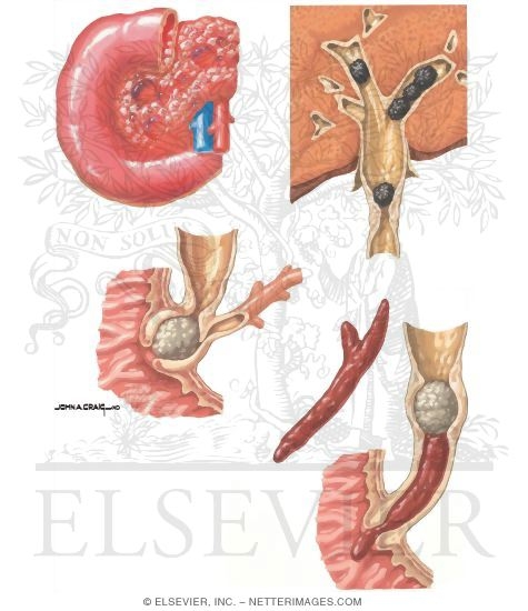
Gall stone impaction occurs at cystic duct.
There's impingement of the gall stone over the common hepatic duct, which eventually results in formation of a fistula in between the gall bladder and the bile duct.
An exception towards the Curvoisier's law.
Complications : Post-cholecystectomy
1) Haemorrhage
Usually the source is from cystic artery.
One should suspect possibility of haemorrhage if the patient complains of persistent abdominal pain, or features of hypovolemic shock during post-operative period
2) Infection
Drastically reduced after administration of prophylactic antibiotics before cholecystectomy.
3) Leakage of bile
Usually due to disrupted ligature, or accidental removal of any accessory ducts.
Patient usually complains of persistent abdominal pain.
If there's no biliary ascites -> ERCP
If there's biliary ascites -> Laparotomy
4) Post-cholecystectomy syndrome
5) Biliary stricture
6) Retained stone
Usually found during T-tube cholangiogram
Any procedure done to explore the common bile duct requires insertion of T-tube.
This is to prevent biliary stasis due to formation of stricture.
The opening of bile duct is sutured to the T-tube, which the long limb is brought out through an abdominal stab incision.
Any bile is collected through a bag.
7-10 days later, T-tube cholangiogram is done (iodine as contrast material)
If there's free flow of contrast material into the duodenum, and there's no residual stone -> remove the T-tube
If there's residual stone :
Stone is small - try using normal saline to irrigate the duct via the T tube (stone eventually migrates to the duodenum)
Stone is large - delayed removal after 4-6 weeks, via radiographically guided removal using dormia basket
Wednesday, May 5, 2010
Tuesday, January 5, 2010
Intercostal drainage
Indications for intercostal drain :
a) Pneumothorax
b) Traumatic haemopneumothorax
c) Malignant pleural effusion
d) Empyema thoracis / Complicated parapneumonic effusion
e) Post-operative drainage : esophagectomy, cardiac surgery, thoracotomy
Equipments :
Intercostal tube or Chest tube (Size for pneumothorax : 36-40 Fr, hemothorax : 22-24 Fr)
Connecting tubes and compatible connectors
Underwater seal drainage bottle containing water upto mark
Line clamp
11 blade scalpel
Instruments for blunt dissection
Blue and Green needle
2 or 3/0 silk in a large hand-held needle
10ml syringe
20ml of 1% lidocaine
Normal saline
Sterile gloves
Sterile drapes
Sterile gauze
Skin prep. solution
Procedures :
1) Explain procedure to the patient if appropriate.
2) Connect the patient to a pulse oxymeter.
3) Prop up the patient to a semi-recumbent position, with the ipsilateral limb abducted.
4) Prepare the skin at the site of tube insertion (antiseptics)
5) Make proper drapping over field of interest.
6) Infiltrate the local anesthetic sufficiently, including the parietal pleura and the periosteum of the rib posterior to the line of incision.
7) Make a transverse, 2cm incison over the 5th intercostal space, over the mid-axillary line (may extend upto the anterior axillary line)
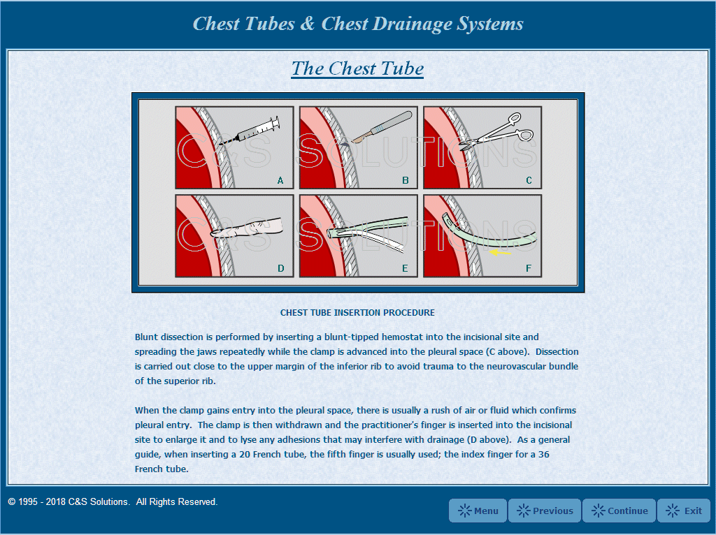
8) Proceed with blunt dissection until the pleura is visible.
9) Now, gently and firmly, by using a blunt-ended clamp, puncture the pleura, and widened the hole created.
10) Place one of your finger into the hole to ensure there's no adhesions.
11) Insert the chest tube without trochar into the puncture hole created after clamping it. Guide the tube superiorly if it's a pneumothorax, and towards the base if it's a hemothorax.
12) Fix the chest tube using the silk sutures.
13) Connect the distal end of the tube to the underwater seal drainage bottle, and remove the clamp.
Potential complications :
1) Misplacement (intra-parenchymal or subcutaneous)
2) Damaging the surrounding structures : liver, spleen, lungs, heart, aorta, diagphram, etc
3) Surgical emphysema
4) Wound infection, empyema
5) Pain
a) Pneumothorax
b) Traumatic haemopneumothorax
c) Malignant pleural effusion
d) Empyema thoracis / Complicated parapneumonic effusion
e) Post-operative drainage : esophagectomy, cardiac surgery, thoracotomy
Equipments :
Intercostal tube or Chest tube (Size for pneumothorax : 36-40 Fr, hemothorax : 22-24 Fr)
Connecting tubes and compatible connectors
Underwater seal drainage bottle containing water upto mark
Line clamp
11 blade scalpel
Instruments for blunt dissection
Blue and Green needle
2 or 3/0 silk in a large hand-held needle
10ml syringe
20ml of 1% lidocaine
Normal saline
Sterile gloves
Sterile drapes
Sterile gauze
Skin prep. solution
Procedures :
1) Explain procedure to the patient if appropriate.
2) Connect the patient to a pulse oxymeter.
3) Prop up the patient to a semi-recumbent position, with the ipsilateral limb abducted.
4) Prepare the skin at the site of tube insertion (antiseptics)
5) Make proper drapping over field of interest.
6) Infiltrate the local anesthetic sufficiently, including the parietal pleura and the periosteum of the rib posterior to the line of incision.
7) Make a transverse, 2cm incison over the 5th intercostal space, over the mid-axillary line (may extend upto the anterior axillary line)
8) Proceed with blunt dissection until the pleura is visible.
9) Now, gently and firmly, by using a blunt-ended clamp, puncture the pleura, and widened the hole created.
10) Place one of your finger into the hole to ensure there's no adhesions.
11) Insert the chest tube without trochar into the puncture hole created after clamping it. Guide the tube superiorly if it's a pneumothorax, and towards the base if it's a hemothorax.
12) Fix the chest tube using the silk sutures.
13) Connect the distal end of the tube to the underwater seal drainage bottle, and remove the clamp.
Potential complications :
1) Misplacement (intra-parenchymal or subcutaneous)
2) Damaging the surrounding structures : liver, spleen, lungs, heart, aorta, diagphram, etc
3) Surgical emphysema
4) Wound infection, empyema
5) Pain
Wednesday, December 30, 2009
Breast Lump
Anatomy of Breast
The vertical extent of breast is from 2nd-6th ribs inclusive.
The horizontal extent is from the lateral edge of sternum to the mid-axillary line.
2/3rds of the breast overlies the pectoralis major muscle, whereas 1/3 of it over the serratus anterior.
The lower medial quadrant is lying on the external oblique aponeurosis, which separates it from the rectus abdominis.
The breast tissue is separated from the pectoralis major muscle by the pectoral fascia. It's anchored anteriorly to the skin, posteriorly to the pectoral fascia by the cooper's ligament.
The outer prolongation of the gland into the axilla at the level of 3rd rib, is known as the axillary tail of spence. It enters the axilla by piercing the opening in the axillary fascia, known as the foramen of langer, and if it's enlarged, it can be mistaken as a lipoma.
The breast tissue is made up of acini, which forms the lobules, and the aggregations of these lobules made up the lobes. Each of these lobes are drained by a collecting duct, and 10-15 of these ducts drains out to the surface of nipple.
If there's a malignant breast lump, infiltrating the cooper's ligament, it'll lead to dimpling of the skin over breast, due to contraction of the cooper's ligament. If the tumour continues to infiltrate along these cooper's ligament, and now involving the pectoral muscle, it renders it lump non-mobile in a direction parallel to the direction of the pectoral muscle fibers, and mobile in a direction perpendicular to it.
If a tumour infiltrates into the major milk ducts, a subsequent fibrosis is going to cause the nipple to be drawn inwards, and hence leading to nipple retraction.
Peu'd orange, an appearance of orange skin of the skin of breast in infiltrative CA breast, is due to the tumour destruction of the cuticle lymphatics, leading to subsequent lymphostasis and edema, and hence the pits of hair follicles appears depressed from the surrounding skin.
Arterial supply
Lateral thoracic artery (major), a branch of the 2nd part of axillary artery
Perforating cutaneous branch of the interal mammary artery to the 2nd, 3rd, 4th space.
Lateral branches of the 2nd, 3rd, 4th intercostal arteries
Venous drainage
Intercostal veins, axillary veins and internal mammary veins
Lymphatic drainage
The primary lymphatic drainage of breast is the axillary nodes (around 20-30 of them), followed by the internal mammary nodes. Around 75% of the lymphatics of the breast is handled by the axillary nodes, and the remaining 25%, by the internal mammary nodes.
There are 5 groups of axillary nodes, namely the anterior, posterior, lateral, central and apical. By surgical means, they can be classified based on their position in relation with the pectoralis minor muscle.
Nodes located below the lateral border the pectoralis minor -> Level I (anterior, posterior and lateral)
Nodes located behind the pectoralis minor muscle -> Level II (central)
Nodes located above the medial border of pectoralis minor muscle -> Level III (apical)
Lymphatics from the lateral quadrant, some from the medial quadrant drains into the anterior nodes (located behind the lower border of pec. major muscle), and the posteior nodes, which then proceeds to the central nodes, and lastly the apical nodes.
Lymphatics from the right axillary and internal mammary nodes drains into the right subclavian lymphatic duct, whilst lymphatics from the left axillary and internal mammary nodes drains into the thoracic duct, then into the subclavian vein. Both eventually drains into the subclavian vein.
Common presenting problem of the breast
1) Painless lump
Breast cancer
Fibroadenoma
An area of fibroadenosis
Breast cyst
2) Painful lump
An area of fibroadenosis
Breast cyst
Periductal mastitis
Breast abscess
Advanced breast carcinoma
3) Only pain
Cyclical mastalgia
Non-cyclical mastalgia
Very rarely, CA breast
4) Nipple changes
Destruction
Depression (retraction, inversion)
Duplication
Discharge
Deviation
Displacement
Remember these 6 Ds
Different causes of nipple discharge :
Fresh red (blood) -> Duct papilloma
Pinkish (blood + serum) -> CA breast
Greenish/Blackish -> Breast cyst
Creamy, pale yellowish -> Duct ectasia
Whitish -> Lactation
Occasionally, paget's disease of the nipple can be confused with eczema of the breast. To differentiate it :
Paget's disease Eczema
Unilateral Bilateral
No vesicles With vesicles
Doesn't itch Itches
May be associated with lump No lump
Nipple may not be intact Nipple is always intact
Post-menopausal Post-lactational
How do you approach in a case of breast lump?
It's by the tripple assessment, which includes history and examination, imaging and Biopsy
1) History
About the lump : Onset, side, site, duration, progression, initial size, current size
Any pain associated with the lump, and proceed to the details of pain
Is there any skin changes? (dimpling, nodules, ulceration, peu'd orange)
Ask about the onset, duration and progression
Is there any nipple discharge?
Ask about the onset, duration, amount, colour, foul-smelling
Is there any recent nipple retraction?
Is there any lumps felt in the axilla?
Then, proceed to the history of risk factors :
Age of menarche (<11 years old)
Age of menopause (>55 years old)
Age of first child birth (if <30 years old, lesser risk)
Parity index (no. of children)
History of breast feeding and the duration (at least 6 months)
Family history of breast cancer (first degree relatives)
HRT/OCP intake (controversial)
Post-menopausal obesity
Diet - Fatty food predilection
Then, h/o of metastases :
Consitutional -> h/o of weight lost, lost of appetite
Respiratory -> Cough, hemoptysis, dyspnoea
CNS -> Headache, vomiting, diplopia, focal neurological deficits, seizures
Liver -> Jaundice
Musculoskeletal -> Bone pain, pathological fractures
2) Examination
a) Comparison of both breasts
Patient is sitting up, both arms are at her side.
Now observe, any discrepency of size and shape of both breasts?
Is there any differences in between the nipples of both sides?
Is there any visible mass?
Now, ask the patient to lift up both of her arms above head
Observe if there's any accentuation of dimpling or distortion of the breasts?
Observe if both breast are elevated equally (if one is higher than the other, it means that the lump probably has fixed to the pec.major muscle)
Now, ask the patient to bend forwards.
Does both breast moves forwards equally?
If one doesn't move as the patient bend forwards, possibly it has fixed to the chest wall (intercostal muscles or ribs)
Now, examine the affected breast.
On inspection, note :
Size and shape - normal?
Skin over breast - peu'd orange, ulcers, nodules, dimpling, dilated veins
Nipple - retraction, discharge
Visible mass - size, shape, surface
Any ulcers - describe it
On palpation, palpate all 4 quadrants of the breast, including the central area and the axillary tail of spence. Note if there's any lump under headings of :
Number of lumps
Site
Size
Surface
Consistency
Tenderness
Edges
Mobility and fixity
First as the patient's hands are placed over her hips, try moving the lump.
If it's not mobile even when the muscles are relaxed, it means that the lump has infiltrated into the chest wall (skin/intercostal muscles).
If it's infiltrated into the serratus anterior, it'll be the same as infiltration to the chest wall, and noted as stage T4 in TMN staging system.
To test whether it has infiltrated to serratus anterior, ask the patient to push against the wall using both hands, and if renders the lump non-mobile, it means infiltration to serratus anterior has taken place.
If the tumour has already infiltrated into the pectoralis major muscle, the lump is mobile in a direction perpendicular to the muscle fibers, but not in a direction parallel to it. This can be confirmed by asking the patient to press firmly using her hands against her hips, and if the lump now is completely immobile, it means infiltration into the pectoralis major muscle has taken place.
Now, try to feel for any lumps of SC nodes.
Examine the axilla, and note any enlarged nodes in it's numbers, consistency, tenderness, fixity.
Percuss the parasternal region for any dullness.
Repeat the same procedure for the opposite breast and axilla.
Now, examine the abdomen -> hepatomegaly, ascites, PR and PV done (metastatic deposits)
Examine the lungs -> Chest wall tenderness, pleural effusions
Check for any bony tenderness
3) Imaging
For women below age of 40 years old, the imaging of choice is ultrasonography
For women above age of 40 years old, imaging of choice is mammography
4) Biopsy
FNAC
TRU-cut/core-needle biopsy
Incisional biopsy
Excisional biopsy
Further test done :
1) Liver function test - elevation of ALP is suggestive of liver metastases
2) Liver ultrasound - liver metastases
3) Chest X ray - pleural effusions, cannon-ball secondaries, rib erosions
4) CT abdomen and Bone scan (optional - not done in MUAR)
TMN staging of CA breast
Tis - Carcinoma in situ
T0 - No evidence of the presence of primary tumour
Tx - Primary tumour cannot be accessed (may be after BCS/mastectomy)
T1 - Size of tumour is < 2cm, not fixed to muslces
T2 - Size of tumour is 2-5cm, fixed to the muscles
T3 - Size of tumour is >5cm
T4a - Involvement of the chest wall
T4b - Involvement of the skin over breast
T4c - Both T4a and T4b present
T4d - Inflammatory carcinoma
N0 - No evidence of nodal metastases clinically
N1 - Ipsilateral axillary nodes palpable, mobile
N2 - Ipsilateral axillary nodes palpable, immobile
N3a - Both infraclavicular and axillary nodes palpable
N3b - Both internal mammary and axillary nodes palpable
N3c - Both axillary and supraclavicular nodes palpable
M0 - No distant metastases
M1 - Distant metastases present
Hence, the stages are :
Stage I - T1 N0 M0
Stage IIA - T0 N1 M0, or T1 N1 M0, or T2 N0 M0
Stage IIB - T2 N1 M0, or T3 N0 M0
Stage IIIA - T0/T1/T2 N2 M0 or T3 N1/N2 M0
Stage IIIB - T4 N0/N1/N2 M0
Stage IIIC - Any T N3 M0
Stage IV - Any T Any N, M1
The vertical extent of breast is from 2nd-6th ribs inclusive.
The horizontal extent is from the lateral edge of sternum to the mid-axillary line.
2/3rds of the breast overlies the pectoralis major muscle, whereas 1/3 of it over the serratus anterior.
The lower medial quadrant is lying on the external oblique aponeurosis, which separates it from the rectus abdominis.
The breast tissue is separated from the pectoralis major muscle by the pectoral fascia. It's anchored anteriorly to the skin, posteriorly to the pectoral fascia by the cooper's ligament.
The outer prolongation of the gland into the axilla at the level of 3rd rib, is known as the axillary tail of spence. It enters the axilla by piercing the opening in the axillary fascia, known as the foramen of langer, and if it's enlarged, it can be mistaken as a lipoma.
The breast tissue is made up of acini, which forms the lobules, and the aggregations of these lobules made up the lobes. Each of these lobes are drained by a collecting duct, and 10-15 of these ducts drains out to the surface of nipple.
If there's a malignant breast lump, infiltrating the cooper's ligament, it'll lead to dimpling of the skin over breast, due to contraction of the cooper's ligament. If the tumour continues to infiltrate along these cooper's ligament, and now involving the pectoral muscle, it renders it lump non-mobile in a direction parallel to the direction of the pectoral muscle fibers, and mobile in a direction perpendicular to it.
If a tumour infiltrates into the major milk ducts, a subsequent fibrosis is going to cause the nipple to be drawn inwards, and hence leading to nipple retraction.
Peu'd orange, an appearance of orange skin of the skin of breast in infiltrative CA breast, is due to the tumour destruction of the cuticle lymphatics, leading to subsequent lymphostasis and edema, and hence the pits of hair follicles appears depressed from the surrounding skin.
Arterial supply
Lateral thoracic artery (major), a branch of the 2nd part of axillary artery
Perforating cutaneous branch of the interal mammary artery to the 2nd, 3rd, 4th space.
Lateral branches of the 2nd, 3rd, 4th intercostal arteries
Venous drainage
Intercostal veins, axillary veins and internal mammary veins
Lymphatic drainage
The primary lymphatic drainage of breast is the axillary nodes (around 20-30 of them), followed by the internal mammary nodes. Around 75% of the lymphatics of the breast is handled by the axillary nodes, and the remaining 25%, by the internal mammary nodes.
There are 5 groups of axillary nodes, namely the anterior, posterior, lateral, central and apical. By surgical means, they can be classified based on their position in relation with the pectoralis minor muscle.
Nodes located below the lateral border the pectoralis minor -> Level I (anterior, posterior and lateral)
Nodes located behind the pectoralis minor muscle -> Level II (central)
Nodes located above the medial border of pectoralis minor muscle -> Level III (apical)
Lymphatics from the lateral quadrant, some from the medial quadrant drains into the anterior nodes (located behind the lower border of pec. major muscle), and the posteior nodes, which then proceeds to the central nodes, and lastly the apical nodes.
Lymphatics from the right axillary and internal mammary nodes drains into the right subclavian lymphatic duct, whilst lymphatics from the left axillary and internal mammary nodes drains into the thoracic duct, then into the subclavian vein. Both eventually drains into the subclavian vein.
Common presenting problem of the breast
1) Painless lump
Breast cancer
Fibroadenoma
An area of fibroadenosis
Breast cyst
2) Painful lump
An area of fibroadenosis
Breast cyst
Periductal mastitis
Breast abscess
Advanced breast carcinoma
3) Only pain
Cyclical mastalgia
Non-cyclical mastalgia
Very rarely, CA breast
4) Nipple changes
Destruction
Depression (retraction, inversion)
Duplication
Discharge
Deviation
Displacement
Remember these 6 Ds
Different causes of nipple discharge :
Fresh red (blood) -> Duct papilloma
Pinkish (blood + serum) -> CA breast
Greenish/Blackish -> Breast cyst
Creamy, pale yellowish -> Duct ectasia
Whitish -> Lactation
Occasionally, paget's disease of the nipple can be confused with eczema of the breast. To differentiate it :
Paget's disease Eczema
Unilateral Bilateral
No vesicles With vesicles
Doesn't itch Itches
May be associated with lump No lump
Nipple may not be intact Nipple is always intact
Post-menopausal Post-lactational
How do you approach in a case of breast lump?
It's by the tripple assessment, which includes history and examination, imaging and Biopsy
1) History
About the lump : Onset, side, site, duration, progression, initial size, current size
Any pain associated with the lump, and proceed to the details of pain
Is there any skin changes? (dimpling, nodules, ulceration, peu'd orange)
Ask about the onset, duration and progression
Is there any nipple discharge?
Ask about the onset, duration, amount, colour, foul-smelling
Is there any recent nipple retraction?
Is there any lumps felt in the axilla?
Then, proceed to the history of risk factors :
Age of menarche (<11 years old)
Age of menopause (>55 years old)
Age of first child birth (if <30 years old, lesser risk)
Parity index (no. of children)
History of breast feeding and the duration (at least 6 months)
Family history of breast cancer (first degree relatives)
HRT/OCP intake (controversial)
Post-menopausal obesity
Diet - Fatty food predilection
Then, h/o of metastases :
Consitutional -> h/o of weight lost, lost of appetite
Respiratory -> Cough, hemoptysis, dyspnoea
CNS -> Headache, vomiting, diplopia, focal neurological deficits, seizures
Liver -> Jaundice
Musculoskeletal -> Bone pain, pathological fractures
2) Examination
a) Comparison of both breasts
Patient is sitting up, both arms are at her side.
Now observe, any discrepency of size and shape of both breasts?
Is there any differences in between the nipples of both sides?
Is there any visible mass?
Now, ask the patient to lift up both of her arms above head
Observe if there's any accentuation of dimpling or distortion of the breasts?
Observe if both breast are elevated equally (if one is higher than the other, it means that the lump probably has fixed to the pec.major muscle)
Now, ask the patient to bend forwards.
Does both breast moves forwards equally?
If one doesn't move as the patient bend forwards, possibly it has fixed to the chest wall (intercostal muscles or ribs)
Now, examine the affected breast.
On inspection, note :
Size and shape - normal?
Skin over breast - peu'd orange, ulcers, nodules, dimpling, dilated veins
Nipple - retraction, discharge
Visible mass - size, shape, surface
Any ulcers - describe it
On palpation, palpate all 4 quadrants of the breast, including the central area and the axillary tail of spence. Note if there's any lump under headings of :
Number of lumps
Site
Size
Surface
Consistency
Tenderness
Edges
Mobility and fixity
First as the patient's hands are placed over her hips, try moving the lump.
If it's not mobile even when the muscles are relaxed, it means that the lump has infiltrated into the chest wall (skin/intercostal muscles).
If it's infiltrated into the serratus anterior, it'll be the same as infiltration to the chest wall, and noted as stage T4 in TMN staging system.
To test whether it has infiltrated to serratus anterior, ask the patient to push against the wall using both hands, and if renders the lump non-mobile, it means infiltration to serratus anterior has taken place.
If the tumour has already infiltrated into the pectoralis major muscle, the lump is mobile in a direction perpendicular to the muscle fibers, but not in a direction parallel to it. This can be confirmed by asking the patient to press firmly using her hands against her hips, and if the lump now is completely immobile, it means infiltration into the pectoralis major muscle has taken place.
Now, try to feel for any lumps of SC nodes.
Examine the axilla, and note any enlarged nodes in it's numbers, consistency, tenderness, fixity.
Percuss the parasternal region for any dullness.
Repeat the same procedure for the opposite breast and axilla.
Now, examine the abdomen -> hepatomegaly, ascites, PR and PV done (metastatic deposits)
Examine the lungs -> Chest wall tenderness, pleural effusions
Check for any bony tenderness
3) Imaging
For women below age of 40 years old, the imaging of choice is ultrasonography
For women above age of 40 years old, imaging of choice is mammography
4) Biopsy
FNAC
TRU-cut/core-needle biopsy
Incisional biopsy
Excisional biopsy
Further test done :
1) Liver function test - elevation of ALP is suggestive of liver metastases
2) Liver ultrasound - liver metastases
3) Chest X ray - pleural effusions, cannon-ball secondaries, rib erosions
4) CT abdomen and Bone scan (optional - not done in MUAR)
TMN staging of CA breast
Tis - Carcinoma in situ
T0 - No evidence of the presence of primary tumour
Tx - Primary tumour cannot be accessed (may be after BCS/mastectomy)
T1 - Size of tumour is < 2cm, not fixed to muslces
T2 - Size of tumour is 2-5cm, fixed to the muscles
T3 - Size of tumour is >5cm
T4a - Involvement of the chest wall
T4b - Involvement of the skin over breast
T4c - Both T4a and T4b present
T4d - Inflammatory carcinoma
N0 - No evidence of nodal metastases clinically
N1 - Ipsilateral axillary nodes palpable, mobile
N2 - Ipsilateral axillary nodes palpable, immobile
N3a - Both infraclavicular and axillary nodes palpable
N3b - Both internal mammary and axillary nodes palpable
N3c - Both axillary and supraclavicular nodes palpable
M0 - No distant metastases
M1 - Distant metastases present
Hence, the stages are :
Stage I - T1 N0 M0
Stage IIA - T0 N1 M0, or T1 N1 M0, or T2 N0 M0
Stage IIB - T2 N1 M0, or T3 N0 M0
Stage IIIA - T0/T1/T2 N2 M0 or T3 N1/N2 M0
Stage IIIB - T4 N0/N1/N2 M0
Stage IIIC - Any T N3 M0
Stage IV - Any T Any N, M1
Tuesday, December 29, 2009
Thoracic Trauma
Introduction
Thoracic trauma accounts for about 25% of all cases of trauma.
Most of the thoracic injuries are life theratening, where the commonest cause of morbidity and mortality is hypoxia and haemorrhage.
However, ironically upto 80% of the cases can be managed conservatively.
The key to succesful management here is early physiological resuscitation and accurate diagnosis.
Investigations
An approach towards chest injuries is the same as any other injuries in primary and secondary survey, as noted by the Advanced Trauma Life Support Protocol (ATLS). History and examination will be important, and probably the most useful tool is a chest radiography.
In an unstable patient, chest radiography can be done first, provided that it didn't interfere with the process of resuscitation. An ultrasound can give useful information about the presence of hematoma together with a contusion or just contusion alone. Chest drain can be both diagnostic and therapeutic, where the benefits outweights the risks.
Some pitfalls during investigations :
a) Failed to identify tracheal shift
b) Failed to pass NG tube due to failure to recognise diagphramatic rupture
c) During hemothorax, must auscultate both anterior and posterior chest
d) Failed to resuscitate the patient first before investigations are done (both should be done hand in hand)
Nowadays, CT scan made an important role in the management of chest injuries.
Not only it can provide details about ribs and verterbral fractures, it can pick up contusions, hematomas, pneumothoraces easily. In penetrating injuries, eg gunshot wounds, CT can even trace the track of penetration through the thorax. Though aortogram is the 'gold standard' in diagnosing disruption of thoracic aorta, CT scan yields the similar results.
Immediately life threatening chest injuries :
a) Airway obstruction
The commonest cause of early preventable death in a case of thoracic injury is airway obstruction, which blood, clots, secretions, dentures, teeth or even tongue can be a source of obstruction. Rapid removal usually relieves the obstruction.
Examples of injuries potentially causing airway obstruction :
a) Expanding neck hematomas
b) Bilateral mandibular fractures
Both a and b causing pharyngeal deviation and tracheal compression
c) Laryngeal injury with thyroid/cricoid cartilage fracture, and other tracheal injuries
What need to be done immediately is endotracheal intubation, as early as possible.
Since most of these conditions are insidious and yet progressive, and delay will render increased difficulty in inserting the ET tube.
b) Tension pneumothorax
Tension pneumothorax occurs when "one-way" valve is created in such a way that air is collected within the pleural cavity, without any means of escape. The source of air leakage can be originating from the chest wall or lung parenchyma. This results in significant compression over the affected lung, obstruction of the great veins compromising the venous return, mediastinal shift and eventually, compression of the opposite lung.
Common causes includes, penetrating chest injuries, blunt chest trauma with parenchymal injury, iatrogenic causes includes a central subclavian venepuncture or mechanical positive pressure ventilation that has gone wrong.
The clinical presentation is dramatic, with a panicky patient, complaints of dyspnoea, and with distended neck veins. Clinical signs : Tracheal shift to the opposite side (late presentation), diminished lung expansion over affected side, hyperresonant note on percussion, absence breath sounds.
Tension pneumothorax is a clinical diagnosis, NEVER EVER proceed to radiological investigations first.
If clinical diagnosis is establish, one should use a large bore needle, puncture the anterior chest and the 2nd intercostal space, along the midclavicular line. This is followed by inserting a chest tube over the 5th intercostal space at the anterior axillary line.
c) Pericardial tamponade
In a case of patient with shock and distended vein, pericardial tamponade must be differentiated from tension pneumothorax. Pericardial tamponade is usually caused by penetrating chest injuries, and due to the non-distensible feature of the pericardial sac, even accumulation of small volume of blood is going to cause significant mechanical obstruction which renders cardiac pump failure.
The typical presentation will be : Features of hemorrhagic shock, Raised JVP and CVP, muffled heart sounds. Some pitfalls of these presentation must be remembered :
i) In case where there's active bleeding from a site distant to site of pathology, the neck veins are not distended.
ii) In case where the patient is having circulatory collapse, CVP will not be raised
To buy time for preparing the patient for definite operative management, which is left thoracotomy and sternotomy, a needle pericardiocentesis and resuscitation can be done. Needle pericardiocentesis is NOT a substitute for surgical management, and is done with ECG guidance (related with high incidence of iatrogenic myocardial injury)
d) Open pneumothorax
This means an opening chest wound is present, where the size of the defect is > 3cm.
Every breath that is inhaled, more air will be accumulated within the affected hemithorax.
This eventually causes significant hypoventilation, and eventually hypoxia.
The signs and symptoms are directly proportional to the size of the defect.
Initial management includes covering the chest wound is a sterile plastic occlusive dressing, which is only adhered at 3 sites, creating a flutter-wave valve, while suction is continued, where the tube is connected to an underwater seal drainage bottle.
Remember, no 'sucking' chest wound should be covered completely before a controlled drainage is established..
Definite management : pulmonary debridement and closure of the wound.
Some pit falls regarding this conditions :
For adults, a larger tube is required (>28 FG in size)
Some patients may require 2 chest drains
In case where patient's condition doesn't improve despite adequate drainage, try reducing the pressure within the seal drainage bottle to 5cm H20.
Early mobilisation and physiotherapy is required
e) Massive hemothorax
Defined by : initial blood collection by chest drain of > 1500 ml or in on-going hemorrhage, > 200-300 ml/h of blood collected over a period of 2-3 hours.
Massive hemothorax usually occurs due to blunt injuries, rupturing the intercostal and internal mammary vessels. Blood is hence collected within the affected hemithorax, causing significant respiratory distress. It's recognised by signs of haemorrhagic shock, flat neck veins, diminished expansion, dullness on percussion, absence of breath sounds.
Initial management of massive hemothorax includes chest drain, resuscitation and sometimes, intubation. Blood from the pleural cavity must be drained as rapid and as complete as possible, in order to prevent possibility of empyema and later, fibrothorax.
Pit falls regarding massive hemothorax :
1) One must examine both anterior and posterior chest when the patient is lying in a supine position, since there's a chance where the affected lung 'floats' within the BLOODY thoracic cavity.
If you only auscultate the anterior chest - it'll be normal
2) Even after draining out about 500ml of blood, dullness still persist and radio-opacity still present -> emergency thoracotomy
f) Flial chest
Flial chest is defined as a loss of bony continuity of a chest wall segment with the rest of thoracic cage, caused by a blunt trauma, which occurs when there's :
i) 3 or more rib fractures
ii) occurs in more than 2-3 places
Flial chest is a clinical diagnosis, not by chest radiography.
It's done by observing few respiratory cycles, where the flial segment will be drawn inwards during inspiration.
Causes of hypoxia in flial chest : voluntary splinting due to pain, pulmonary contusion, defect in the mechanical movement of the rib cage
Initial management : opiate analgesics, oxygen support. If a chest drain is present, intrapleural local analgesia can be given. Ventilation is reseved for patients with respiratory failure despite optimal treatment given. Surgical fixation is done in severe thoracic injury or in cases where pulmonary contusion is present.
Thoracic trauma accounts for about 25% of all cases of trauma.
Most of the thoracic injuries are life theratening, where the commonest cause of morbidity and mortality is hypoxia and haemorrhage.
However, ironically upto 80% of the cases can be managed conservatively.
The key to succesful management here is early physiological resuscitation and accurate diagnosis.
Investigations
An approach towards chest injuries is the same as any other injuries in primary and secondary survey, as noted by the Advanced Trauma Life Support Protocol (ATLS). History and examination will be important, and probably the most useful tool is a chest radiography.
In an unstable patient, chest radiography can be done first, provided that it didn't interfere with the process of resuscitation. An ultrasound can give useful information about the presence of hematoma together with a contusion or just contusion alone. Chest drain can be both diagnostic and therapeutic, where the benefits outweights the risks.
Some pitfalls during investigations :
a) Failed to identify tracheal shift
b) Failed to pass NG tube due to failure to recognise diagphramatic rupture
c) During hemothorax, must auscultate both anterior and posterior chest
d) Failed to resuscitate the patient first before investigations are done (both should be done hand in hand)
Nowadays, CT scan made an important role in the management of chest injuries.
Not only it can provide details about ribs and verterbral fractures, it can pick up contusions, hematomas, pneumothoraces easily. In penetrating injuries, eg gunshot wounds, CT can even trace the track of penetration through the thorax. Though aortogram is the 'gold standard' in diagnosing disruption of thoracic aorta, CT scan yields the similar results.
Immediately life threatening chest injuries :
a) Airway obstruction
The commonest cause of early preventable death in a case of thoracic injury is airway obstruction, which blood, clots, secretions, dentures, teeth or even tongue can be a source of obstruction. Rapid removal usually relieves the obstruction.
Examples of injuries potentially causing airway obstruction :
a) Expanding neck hematomas
b) Bilateral mandibular fractures
Both a and b causing pharyngeal deviation and tracheal compression
c) Laryngeal injury with thyroid/cricoid cartilage fracture, and other tracheal injuries
What need to be done immediately is endotracheal intubation, as early as possible.
Since most of these conditions are insidious and yet progressive, and delay will render increased difficulty in inserting the ET tube.
b) Tension pneumothorax
Tension pneumothorax occurs when "one-way" valve is created in such a way that air is collected within the pleural cavity, without any means of escape. The source of air leakage can be originating from the chest wall or lung parenchyma. This results in significant compression over the affected lung, obstruction of the great veins compromising the venous return, mediastinal shift and eventually, compression of the opposite lung.
Common causes includes, penetrating chest injuries, blunt chest trauma with parenchymal injury, iatrogenic causes includes a central subclavian venepuncture or mechanical positive pressure ventilation that has gone wrong.
The clinical presentation is dramatic, with a panicky patient, complaints of dyspnoea, and with distended neck veins. Clinical signs : Tracheal shift to the opposite side (late presentation), diminished lung expansion over affected side, hyperresonant note on percussion, absence breath sounds.
Tension pneumothorax is a clinical diagnosis, NEVER EVER proceed to radiological investigations first.
If clinical diagnosis is establish, one should use a large bore needle, puncture the anterior chest and the 2nd intercostal space, along the midclavicular line. This is followed by inserting a chest tube over the 5th intercostal space at the anterior axillary line.
c) Pericardial tamponade
In a case of patient with shock and distended vein, pericardial tamponade must be differentiated from tension pneumothorax. Pericardial tamponade is usually caused by penetrating chest injuries, and due to the non-distensible feature of the pericardial sac, even accumulation of small volume of blood is going to cause significant mechanical obstruction which renders cardiac pump failure.
The typical presentation will be : Features of hemorrhagic shock, Raised JVP and CVP, muffled heart sounds. Some pitfalls of these presentation must be remembered :
i) In case where there's active bleeding from a site distant to site of pathology, the neck veins are not distended.
ii) In case where the patient is having circulatory collapse, CVP will not be raised
To buy time for preparing the patient for definite operative management, which is left thoracotomy and sternotomy, a needle pericardiocentesis and resuscitation can be done. Needle pericardiocentesis is NOT a substitute for surgical management, and is done with ECG guidance (related with high incidence of iatrogenic myocardial injury)
d) Open pneumothorax
This means an opening chest wound is present, where the size of the defect is > 3cm.
Every breath that is inhaled, more air will be accumulated within the affected hemithorax.
This eventually causes significant hypoventilation, and eventually hypoxia.
The signs and symptoms are directly proportional to the size of the defect.
Initial management includes covering the chest wound is a sterile plastic occlusive dressing, which is only adhered at 3 sites, creating a flutter-wave valve, while suction is continued, where the tube is connected to an underwater seal drainage bottle.
Remember, no 'sucking' chest wound should be covered completely before a controlled drainage is established..
Definite management : pulmonary debridement and closure of the wound.
Some pit falls regarding this conditions :
For adults, a larger tube is required (>28 FG in size)
Some patients may require 2 chest drains
In case where patient's condition doesn't improve despite adequate drainage, try reducing the pressure within the seal drainage bottle to 5cm H20.
Early mobilisation and physiotherapy is required
e) Massive hemothorax
Defined by : initial blood collection by chest drain of > 1500 ml or in on-going hemorrhage, > 200-300 ml/h of blood collected over a period of 2-3 hours.
Massive hemothorax usually occurs due to blunt injuries, rupturing the intercostal and internal mammary vessels. Blood is hence collected within the affected hemithorax, causing significant respiratory distress. It's recognised by signs of haemorrhagic shock, flat neck veins, diminished expansion, dullness on percussion, absence of breath sounds.
Initial management of massive hemothorax includes chest drain, resuscitation and sometimes, intubation. Blood from the pleural cavity must be drained as rapid and as complete as possible, in order to prevent possibility of empyema and later, fibrothorax.
Pit falls regarding massive hemothorax :
1) One must examine both anterior and posterior chest when the patient is lying in a supine position, since there's a chance where the affected lung 'floats' within the BLOODY thoracic cavity.
If you only auscultate the anterior chest - it'll be normal
2) Even after draining out about 500ml of blood, dullness still persist and radio-opacity still present -> emergency thoracotomy
f) Flial chest
Flial chest is defined as a loss of bony continuity of a chest wall segment with the rest of thoracic cage, caused by a blunt trauma, which occurs when there's :
i) 3 or more rib fractures
ii) occurs in more than 2-3 places
Flial chest is a clinical diagnosis, not by chest radiography.
It's done by observing few respiratory cycles, where the flial segment will be drawn inwards during inspiration.
Causes of hypoxia in flial chest : voluntary splinting due to pain, pulmonary contusion, defect in the mechanical movement of the rib cage
Initial management : opiate analgesics, oxygen support. If a chest drain is present, intrapleural local analgesia can be given. Ventilation is reseved for patients with respiratory failure despite optimal treatment given. Surgical fixation is done in severe thoracic injury or in cases where pulmonary contusion is present.
Saturday, December 26, 2009
A case of Acute testicular pain
History
We have a 16 years old male here presented to the ER complaining of sudden onset of right testicular pain. The pain woke him up from his sleep and has persisted over the last 3 hrs. His mother says that he has vomited once. His previous medical history includes a similar event a year ago, but on that occasion the pain subsided quickly. He is an asthmatic and uses a salbutamol inhaler.
Only with h/o, what's your differential diagnosis?
Testicular torsion?
Acute epididymo-orchitis?
Torsion of appendix testis?
Infected hydrocele?
Strangulated hernia?
Testicular rupture?
Haemorrhage into a tumour?
On examination
The left hemi-scrotum feels normal but the right side is acutely swollen and tender on palpation. The testicle is elevated when compared to the other side and has an abnormal horizontal lie. The abdomen is soft, non tender, with intact hernial orifices. Vitals are stable, cremesteric reflex is absent.
So, what's your provisional diagnosis?
In this case, testicular torsion should be ruled out unless proven otherwise. Points towards diagnosis of testicular torsion :
1) Age (testicular torsion is common in age group of 10-25 yrs old)
2) Elevated, tender right testicle
3) Abnormal horizontal lie (risk factor for torsion)
4) Cremesteric reflex is absent (bear in mind that presence of this reflex doesn't rule out testicular torsion!)
If doppler's ultrasound is immediately available, a results showing interrupted blood supply to the testis is diagnostic.
However, if the diagnosis is in doubt, PLS peform surgical exploration to confirm the diagnosis. If not, he CAN SUE YOU BECOZ you've caused him to lose his precious balls.
Remember, you've only 4-6 hours (starting from the time of onset of pain) to salvage the balls.
However, if the patient presented within the first hour after onset of pain, it's sometimes possible to untwist the cord manually, which if succesful, the affected testicle is out of danger and surgery can be planned later.
And, surgical correction is bilateral, since congenital defects often involves both sides.
We have a 16 years old male here presented to the ER complaining of sudden onset of right testicular pain. The pain woke him up from his sleep and has persisted over the last 3 hrs. His mother says that he has vomited once. His previous medical history includes a similar event a year ago, but on that occasion the pain subsided quickly. He is an asthmatic and uses a salbutamol inhaler.
Only with h/o, what's your differential diagnosis?
Testicular torsion?
Acute epididymo-orchitis?
Torsion of appendix testis?
Infected hydrocele?
Strangulated hernia?
Testicular rupture?
Haemorrhage into a tumour?
On examination
The left hemi-scrotum feels normal but the right side is acutely swollen and tender on palpation. The testicle is elevated when compared to the other side and has an abnormal horizontal lie. The abdomen is soft, non tender, with intact hernial orifices. Vitals are stable, cremesteric reflex is absent.
So, what's your provisional diagnosis?
In this case, testicular torsion should be ruled out unless proven otherwise. Points towards diagnosis of testicular torsion :
1) Age (testicular torsion is common in age group of 10-25 yrs old)
2) Elevated, tender right testicle
3) Abnormal horizontal lie (risk factor for torsion)
4) Cremesteric reflex is absent (bear in mind that presence of this reflex doesn't rule out testicular torsion!)
If doppler's ultrasound is immediately available, a results showing interrupted blood supply to the testis is diagnostic.
However, if the diagnosis is in doubt, PLS peform surgical exploration to confirm the diagnosis. If not, he CAN SUE YOU BECOZ you've caused him to lose his precious balls.
Remember, you've only 4-6 hours (starting from the time of onset of pain) to salvage the balls.
However, if the patient presented within the first hour after onset of pain, it's sometimes possible to untwist the cord manually, which if succesful, the affected testicle is out of danger and surgery can be planned later.
And, surgical correction is bilateral, since congenital defects often involves both sides.
Tuesday, December 15, 2009
Head injury - part 3
Management of mild head injury (GCS 14-15)
Most of the occasions, patients with mild head injury, after history and examination, and a period of observation, will be allowed to be discharge after following criterias met :
Battle's sign
a) Full GCS score (15/15)
b) No focal neurological deficits
c) Accompanied by a responsible adult
d) Not under influence of any drugs/alcohol
e) Verbal/Written advice about the injury given
Statement e) means : Advice regarding any worsening of symptoms, such as persistent headahce not relieved by analgesia, severe vomiting, blurring of vision, diplopia, weakness/numbness of limbs have been given verbally or written.
Sometimes, for patients with mild head injury, decision of whether to perform CT brain or not can be a big headahce. However, here are the NICE guidelines regarding indications of CT brain in patients with mild head injury :
a) GCS is <13 at any point
b) GCS is 13-14 at 2 hours time
c) Evidence of focal neurological deficit
d) Suspicion of open, comminuted, depressed, or basal skull fracture
e) Vomiting > 1 episode
f) Seizures
Urgent indication
a) Age > 65 years old
b) Evidence of coagulopathy (liver disease, blood dyscarias, warfarin, anti-platelet medications)
c) Dangerous mechanism of head injury (CT within 8 hrs)
d) Antegrade amnesia > 30 mins (CT within 8 hrs)
Management of moderate/severe head injury
First of all, resuscitation and primary survery.
After stabilising cervical spine at 3 fixation point, start primary surveying.
Remember that normalising the patient's oxygenation and circulation is more important than getting a CT done! This is to prevent secondary brain injury
After primary survey, you've made a diagnosis of moderate/severe head injury, the next step is CT brain, to detect any intracranial hematoma, or any skull fractures, soft tissue injuries, or any mild intracerebral contusion.
For intubated patients, it's recommended that you've asked for CT cervical spine.
Before ariving at the hospital, some conservative management can be given for raised ICP, which includes :
a) Reversed tredelenburg : Raised head upto 20-30 degrees
b) Check if the cervical collar is too tight (may obstruct venous drainage from brain)
c) If there's pupillary dilatation (may be due to acute raised ICP), 0.5mg/kg 20% IV mannitol can be given.
Medical management of severe head injury
Severe head injury is preferably managed in a neurointensive care unit.
ICP can be monitored by passing a catheter into the frontal horn of the lateral ventricle (2 finger breadth from the blurred hole, behind the hairline)
Raise the patient's head for about 20-30 degrees
Protect the patient's airway!
For those with traumatic brain injury and coma, they are more prone to aspiration.
Preferably intubate the patient, and provide high flow oxygen. (Prevent hypoxia)
Make sure that the cervical collar is not too tight.
Cerebral vasculatures are very sensitive to the PCo2 level. When there's a rise in PCo2 level, the cerebral vasculatures dilates, and elevates the ICP. In contrast, when there's a fall in PCo2 level, cerebral vasculature constricts.
Hence, you must try to maintain the PCo2 level in between 4.5-5kPa.
Some experienced anesthetist may induce hyperventilation in patients to cause temporary reduction in ICP by reducing the PCo2 level.
Sedative given, either with or without muscle relaxant.
Mannitol/Frusemide given to reduce cerebral edema.
Patient is prone for hyponatremia or other electrolyte imbalance -> correct it
Avoid pyrexia, as it'll cause undesirable increase in the brain metabolic activity.
Barbiturates eg: thiopentone sodium is given to reduce ICP and brain metabolic rate.
Prophylactic anticonvulsant given.
Most of the occasions, patients with mild head injury, after history and examination, and a period of observation, will be allowed to be discharge after following criterias met :
Battle's sign
a) Full GCS score (15/15)
b) No focal neurological deficits
c) Accompanied by a responsible adult
d) Not under influence of any drugs/alcohol
e) Verbal/Written advice about the injury given
Racoon's Sign
Statement e) means : Advice regarding any worsening of symptoms, such as persistent headahce not relieved by analgesia, severe vomiting, blurring of vision, diplopia, weakness/numbness of limbs have been given verbally or written.
Sometimes, for patients with mild head injury, decision of whether to perform CT brain or not can be a big headahce. However, here are the NICE guidelines regarding indications of CT brain in patients with mild head injury :
a) GCS is <13 at any point
b) GCS is 13-14 at 2 hours time
c) Evidence of focal neurological deficit
d) Suspicion of open, comminuted, depressed, or basal skull fracture
e) Vomiting > 1 episode
f) Seizures
Urgent indication
a) Age > 65 years old
b) Evidence of coagulopathy (liver disease, blood dyscarias, warfarin, anti-platelet medications)
c) Dangerous mechanism of head injury (CT within 8 hrs)
d) Antegrade amnesia > 30 mins (CT within 8 hrs)
Management of moderate/severe head injury
First of all, resuscitation and primary survery.
After stabilising cervical spine at 3 fixation point, start primary surveying.
Remember that normalising the patient's oxygenation and circulation is more important than getting a CT done! This is to prevent secondary brain injury
After primary survey, you've made a diagnosis of moderate/severe head injury, the next step is CT brain, to detect any intracranial hematoma, or any skull fractures, soft tissue injuries, or any mild intracerebral contusion.
For intubated patients, it's recommended that you've asked for CT cervical spine.
Before ariving at the hospital, some conservative management can be given for raised ICP, which includes :
a) Reversed tredelenburg : Raised head upto 20-30 degrees
b) Check if the cervical collar is too tight (may obstruct venous drainage from brain)
c) If there's pupillary dilatation (may be due to acute raised ICP), 0.5mg/kg 20% IV mannitol can be given.
Medical management of severe head injury
Severe head injury is preferably managed in a neurointensive care unit.
ICP can be monitored by passing a catheter into the frontal horn of the lateral ventricle (2 finger breadth from the blurred hole, behind the hairline)
Raise the patient's head for about 20-30 degrees
Protect the patient's airway!
For those with traumatic brain injury and coma, they are more prone to aspiration.
Preferably intubate the patient, and provide high flow oxygen. (Prevent hypoxia)
Make sure that the cervical collar is not too tight.
Cerebral vasculatures are very sensitive to the PCo2 level. When there's a rise in PCo2 level, the cerebral vasculatures dilates, and elevates the ICP. In contrast, when there's a fall in PCo2 level, cerebral vasculature constricts.
Hence, you must try to maintain the PCo2 level in between 4.5-5kPa.
Some experienced anesthetist may induce hyperventilation in patients to cause temporary reduction in ICP by reducing the PCo2 level.
Sedative given, either with or without muscle relaxant.
Mannitol/Frusemide given to reduce cerebral edema.
Patient is prone for hyponatremia or other electrolyte imbalance -> correct it
Avoid pyrexia, as it'll cause undesirable increase in the brain metabolic activity.
Barbiturates eg: thiopentone sodium is given to reduce ICP and brain metabolic rate.
Prophylactic anticonvulsant given.
Head Injury - part 2
Extradural hematoma
This refers to collection of blood in between the skull and dura mater.
More commonly seen in younger patients (children, adolescence)
Extradural hematoma is always associated with skull fractures, most frequently, the temporal bone. (since pterion is the thinnest part of skull, involvement of this area causes tearing of the middle meningeal artery)
Of course, involvement of the posterior fossa and frontal bone is also possible.
However, the hematoma is not always arterial in origin, it may be due to a tear to the dural venous sinuses as well.
Classical presentation of extradural hematoma is : (<1/3 of the cases)
Lucid interval, where after initial injury, patient is conscious, alert, oriented, and only complaints of headache. Minutes or hours later, the condition worsens, with deterioration of consciousness, contralateral hemiparesis/plegia, and ipsilateral pupillary dilatation.
Early diagnosis and treatment of subdural hematoma is VITAL.
CT brain is confirmatory, where it'll appears as a lentiform, biconvex, or lense-shaped hyperdense mass in between the skull and brain, with or without midline shift.
After diagnosis is confirmed, surgical evacuation of the hematoma is required, where craniotomy is performed.
Acute subdural hematoma (ASH)
This is actually more common, with poorer prognosis, higher mortality rate as compared to extradural hematoma.
It refers to blood collection in between the dura and arachnoid mater.
ASH is almost always associated with a primary brain injury.
Most of the time at presentation, the patient has impaired consciousness, which rapidly deteriorates depending on the size of the hematoma.
Again, CT brain is diagnostic.
It'll appears as a crescent shaped, more diffuse (with concavity towards the brain), hyperdense mass in between the brain and skull.
Treatment - surgical evacuation by craniotomy
Chronic subdural hematoma (CSH)
CSH often seen in elderly patients, who is on anti-platelets or anti-coagulants. It is believed to be due to tearing of the bridging veins, which causes formation of clinically inapparent, small ASH. Later, as it breaks down and the volume expands, it becomes symptommatic.
Mostly, patients presents with headache, focal neurological deficit, impaired cognition, seizures, etc (hence, one of the d/d of CVA)
CT brain intepretation :
Acute blood (0-10 days) = hyperdense
Subacute blood (10 days - 2 weeks) = isodense
Chronic blood (>2weeks) = hypodense
Treatment = creating a blurr hole and evacuate the hematoma
This refers to collection of blood in between the skull and dura mater.
More commonly seen in younger patients (children, adolescence)
Extradural hematoma is always associated with skull fractures, most frequently, the temporal bone. (since pterion is the thinnest part of skull, involvement of this area causes tearing of the middle meningeal artery)
Of course, involvement of the posterior fossa and frontal bone is also possible.
However, the hematoma is not always arterial in origin, it may be due to a tear to the dural venous sinuses as well.
Classical presentation of extradural hematoma is : (<1/3 of the cases)
Lucid interval, where after initial injury, patient is conscious, alert, oriented, and only complaints of headache. Minutes or hours later, the condition worsens, with deterioration of consciousness, contralateral hemiparesis/plegia, and ipsilateral pupillary dilatation.
Early diagnosis and treatment of subdural hematoma is VITAL.
CT brain is confirmatory, where it'll appears as a lentiform, biconvex, or lense-shaped hyperdense mass in between the skull and brain, with or without midline shift.
After diagnosis is confirmed, surgical evacuation of the hematoma is required, where craniotomy is performed.
Acute subdural hematoma (ASH)
This is actually more common, with poorer prognosis, higher mortality rate as compared to extradural hematoma.
It refers to blood collection in between the dura and arachnoid mater.
ASH is almost always associated with a primary brain injury.
Most of the time at presentation, the patient has impaired consciousness, which rapidly deteriorates depending on the size of the hematoma.
Again, CT brain is diagnostic.
It'll appears as a crescent shaped, more diffuse (with concavity towards the brain), hyperdense mass in between the brain and skull.
Treatment - surgical evacuation by craniotomy
Chronic subdural hematoma (CSH)
CSH often seen in elderly patients, who is on anti-platelets or anti-coagulants. It is believed to be due to tearing of the bridging veins, which causes formation of clinically inapparent, small ASH. Later, as it breaks down and the volume expands, it becomes symptommatic.
Mostly, patients presents with headache, focal neurological deficit, impaired cognition, seizures, etc (hence, one of the d/d of CVA)
CT brain intepretation :
Acute blood (0-10 days) = hyperdense
Subacute blood (10 days - 2 weeks) = isodense
Chronic blood (>2weeks) = hypodense
Treatment = creating a blurr hole and evacuate the hematoma
Subscribe to:
Posts (Atom)